
Explainer · August 7, 2026 · 5 min · By Xander Beaumont
Gland or Fat? Why the Pinch Test Decides Whether Liposuction Alone Will Work
True gynecomastia and pseudogynecomastia look similar in the mirror but respond very differently to treatment. Here is how clinicians tell them apart, and why getting the diagnosis right before surgery matters more than the technique itself.
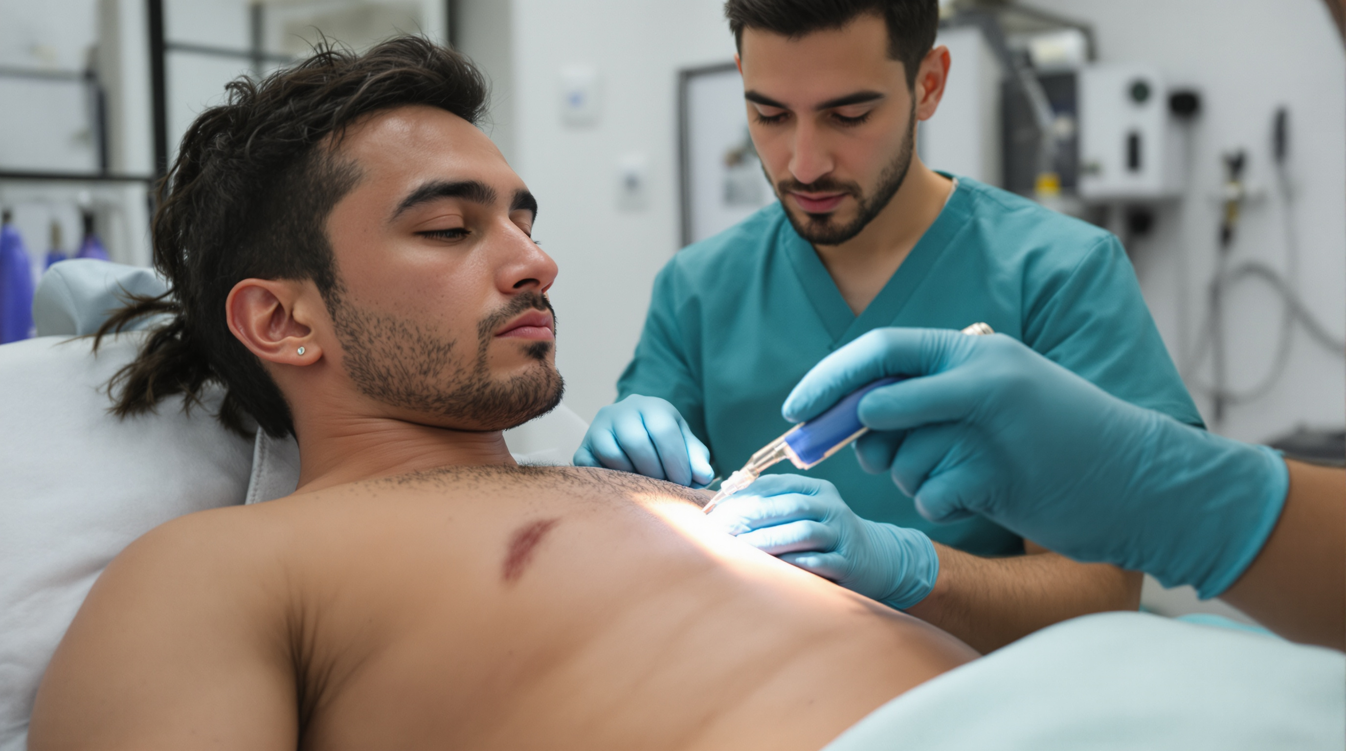
One of the most common disappointments in male chest contouring follows a predictable script. A patient undergoes liposuction of the chest, the swelling resolves over several weeks, and a firm mound remains directly behind the nipple. The fat is gone, but the shape barely changed. In most of these cases the problem was not the surgeon's technique. It was the diagnosis.
Two conditions, one silhouette. True gynecomastia is the growth of glandular breast tissue in men, a dense, fibrous structure driven by hormonal signaling, most often an imbalance in the ratio of estrogen activity to androgen activity at the breast tissue level. Pseudogynecomastia, sometimes called lipomastia, is simply excess subcutaneous fat over the pectoral region with no meaningful glandular component. From across a room the two can look identical. Under the skin they are entirely different materials, and that difference determines what a cannula can and cannot remove.
Why the distinction changes everything. Liposuction works by mechanically disrupting and aspirating adipose tissue, which is soft and loosely organized. Glandular breast tissue is not adipose. It is a firm, rubbery matrix of ducts and fibrous stroma that resists suction the way cartilage resists a straw. Standard liposuction passes around it. Even energy assisted methods, such as ultrasound assisted or power assisted liposuction, thin the fat surrounding the gland but rarely eliminate a well developed glandular disc. When gland is present, most surgeons combine liposuction with direct excision, typically through a small incision along the lower border of the areola, to physically remove the firm tissue. Skipping that step in a true gynecomastia case is the single most common reason for revision surgery in this field.
How clinicians actually check. The core maneuver is straightforward and has been taught for decades. With the patient lying flat and hands behind the head, the examiner places the thumb and forefinger on opposite sides of the chest and slowly brings them together toward the nipple. In true gynecomastia, the fingers meet a discrete, rubbery, often slightly tender disc of tissue centered under the areola, usually at least a couple of centimeters across, and it feels distinct from the softer fat around it. In pseudogynecomastia, the fingers glide through uniform soft tissue until they nearly touch, with no ridge or disc. The gland is also typically concentric around the nipple, while fat distributes more diffusely across the whole chest and often extends toward the armpit.
Imaging is not routine but has a role. Ultrasound can confirm glandular tissue when the exam is ambiguous, and it becomes important when anything atypical appears: a hard, fixed, or one sided mass, nipple discharge, skin changes, or enlarged lymph nodes. Male breast cancer is rare but real, and unilateral firm masses in older men deserve evaluation before any cosmetic procedure is discussed.
The overlap case is the usual case. In practice, most men presenting for chest contouring have a mixture: some gland, some fat, in varying proportions. Adolescent onset gynecomastia that persisted into adulthood tends to be gland dominant. Weight gain related chest fullness in middle age tends to be fat dominant, though years of elevated body fat can itself promote glandular growth, because fat tissue contains aromatase, the enzyme that converts testosterone to estrogen. That mechanism explains a frustrating clinical reality: significant weight loss often shrinks the chest but leaves a residual firm mound, essentially unmasking the gland that was hiding inside the fat.
What patients can reasonably do first. For fat dominant presentations, weight loss genuinely helps and should generally precede surgery, both for results and safety. For gland dominant tissue, no amount of diet, cardio, or pectoral training will remove it, because exercise does not shrink fibrous glandular stroma. Building the pectoral muscle can even push the gland forward and make it more visible. It is also worth reviewing medications and supplements with a physician, since anabolic steroids, certain acid reflux and psychiatric medications, and some prostate treatments are documented contributors. New onset gynecomastia in an adult warrants a medical workup before a surgical conversation, because it can occasionally signal a hormonal or testicular problem.
Questions worth asking at a consultation. Ask directly whether the examiner felt glandular tissue and, if so, whether the plan includes excision or relies on liposuction alone. Ask how residual gland would be handled if found during surgery. Ask about skin quality, because after major weight loss the limiting factor may be skin elasticity rather than tissue volume, and that opens a separate discussion about skin tightening or excision patterns.
The takeaway is unglamorous but reliable: the physical exam done in the first ten minutes of a consultation predicts the surgical outcome better than any device marketing. Fat responds to suction. Gland responds to excision. A plan that matches the tissue is the entire game.